- Talocrural Joint (Ankle Joint)
- Subtalar Joint
- Inferior Tibiofibular Joint
All of these combine to produce the main foot and ankle movements of:
- Plantarflexion (Toes Down)
- Dorsiflexion (Toes Up)
- Inversion (Tilt In)
- Eversion (Tilt Out)
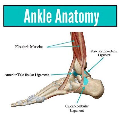
The ankle ligaments criss-cross around these joints to stabilize them at the end of their range of motion, during activities like cutting, jumping, hopping. The main ligaments are:
- Anterior Talofibular Ligament
- Posterior Talofibular Ligament
- Calcaneofibular Ligament
These are probably the most common sprained ligaments in the body! Whether one, two or more of these are stretched determines injury severity. I also included the fibularis muscles . This is because they’re also crucial stabilizers of the ankle. They prevent excessive inversion by attaching from the side of the leg, to under the foot (a natural stirrup). They’re commonly damaged when rolling the ankle, meaning ankle injuries actually damage muscles and tendons as well. Stay tuned for next post for ankle rehab!


